Brain tumours
The majority of adult tumours are supratentorial, where as the majority of childhood tumours are infratentorial.
| Type of tumour | Features |
|---|---|
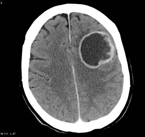
| Gliolastoma multiforme | • The most common primary brain tumour in adults. • Histology: Pleomorphic tumour cells border necrotic areas |
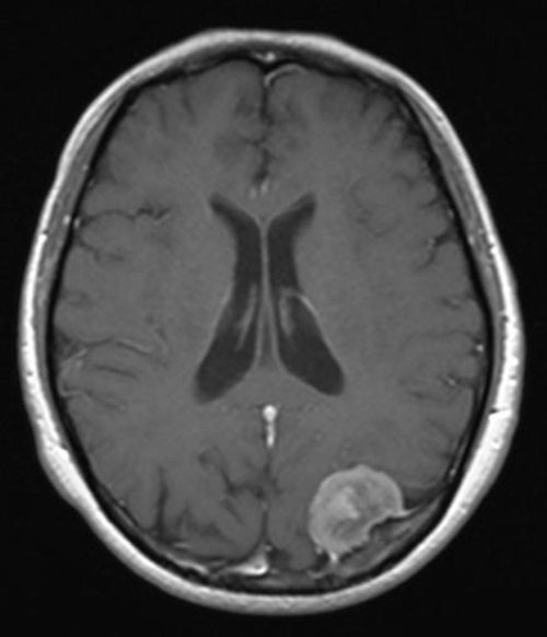
| Meningioma | • The second most common primary brain tumour in adults • Histology: Spindle cells in concentric whorls and calcified psammoma bodies |
| Schwannoma | • Often seen in the cerebellopontine angle: acoustic neuroma • Bilateral schwannoms are seen in neurofibromatosis • Histology: Antoni A or B patterns are seen. Verocay bodies (acellular areas surrounded by nuclear palisades) |
| Pilocytic astrocytoma | • The most common primary brain tumour in children • Histology: Rosenthal fibres (corkscrew eosinophilic bundle) |
| Medulloblastoma | • More common in children • Found exclusively in the posterior fossa • Metatases through the CSF • Histology: Small, blue cells. Rosette pattern of cells with many mitotic figures |
| Ependymoma | • Commonly seen in the 4th ventricle • May cause hydrocephalus • Histology: perivascular pseudorosettes |
| Oligodendroma | • Benign, slow-growing tumour common in the frontal lobes • Histology: Calcifications with 'fried-egg' appearance |
| Haemangioblastoma | • Vascular tumour of the cerebellum • Associated with von Hippel-Lindau syndrome • Histology: foam cells and high vascularity |
| Pituitary adenoma | • Most common type is a prolactinoma • May present with bitemporal hemianopia |
| Craniopharyngioma | • Most common paediatric supratentorial tumour • Histology: Derived from remnants of Rathke pouch |
| Metastases | • Most common type of brain tumour |
 | |
| © Image used on license from Radiopaedia |  |
Meningioma - MRI showing the typical well-circumscribed appearance. A dural tail can be where the tumour 'connects' to the dura. It is seen in around 65% of meningiomas.
 | |
| © Image used on license from Radiopaedia |  |
Glioblastoma multiforme - CT showing a peripherally enhancing lesion within the left frontal lobe. Note the contrast to the more homogenous meningioma above.
