Showing posts with label Notes : Neuroscience. Show all posts

5-HIAA

5-HIAA


During the 1980's there was brief interest in a serotonin metabolite called 5-hydroxyindoleacetic acid (5-HIAA). It was found to have low concentrations in the CSF of up to a third of people with depression and very few normal controls.

People with low CSF levels of 5-HIAA have also been found to respond less well to antidepressants and also to be more likely to commit suicide (this is a well replicated finding).

Low levels of 5-HIAA are also associated with increased levels of aggression

Acoustic neuroma

Acoustic neuroma


Acoustic neuromas (more correctly called vestibular schwannomas) account for approximately five percent of intracranial tumours and 90 percent of cerebellopontine angle

Features can be predicted by the affected cranial nerves
  • cranial nerve VIII: hearing loss, vertigo, tinnitus
  • cranial nerve V: absent corneal reflex
  • cranial nerve VII: facial palsy

Bilateral acoustic neuromas are seen in neurofibromatosis type 2

MRI of the cerebellopontine angle is the investigation of choice

Action potentials

Action potentials


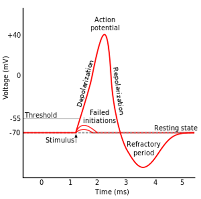
Most cells have a membrane potential that exists across the plasma membrane. In neurons and muscle cells this membrane potential can change over time. When a cell is not stimulated, it is in a resting state and the inside of the cell is negatively charged with respect to the outside. 

The membrane potential of the resting state is -70mV. This negative charge is due to a high concentration of Na+ outside compared to the K+ inside the cell. This ionic gradient is maintained by the Na/K pump.


For an action potential to take place first the membrane potential must be raised to about -55mV. This is done by a neurotransmitter binding to the postsynaptic neuron and opening some ion channels. Once it reaches -55mV it initiates an irreversible process whereby a large number of Na channels open and cause the cell to depolarise. Once the membrane potential reaches about +40 the Na channels close and the K gates open, the K floods out and the membrane potential falls back again

Agnosia

Agnosia


Agnosia refers to loss of ability to recognize objects, persons, sounds, shapes, or smells while the specific sense is not defective nor is there any significant memory loss.

ProsopagnosiaInability to recognise familiar faces
AnosognosiaInability to recognise own condition/ illness
AutotopagnosiaInability to orient parts of the body
PhonagnosiaInability to recognize familiar voices
SimultanagnosiaInability to appreciate two objects in the visual field at the same time
AstereoagnosiaInability to recognize objects by touch

Alexia without agraphia

Alexia without agraphia


In alexia without agraphia the patient cannot read (alexia) but is able to write. Understanding spoken language and conversation are intact.

This is usually due to a lesion destroying the left visual cortex, as well as the connections to the right visual cortex in the corpus callosum. It is typically caused by an occlusion of a branch of the PCA

Alpha-secretase

Alpha-secretase


Alpha-secretase is hoped to offer a new way of treating/ preventing Alzheimers disease.

Once amyloid precursor protein (APP) crosses the cell membrane it is cleaved by a number of different enzymes. Alpha-secretase cleaves the protein in such a way as to produce protein fragments that are non-toxic. In addition to alpha-secretase, there are two other enzymes in the cell that can also cleave APP (beta and gamma-secretase). When these cleave APP the result is shorter, stickier fragments called beta-amyloid, which join together to produce the insoluble amyloid plaques.

Drugs are being developed which either stimulate alpha-secretase or block beta- and gamma-secretase

Amygdala

Amygdala


The amygdala is an olive shaped structure located medially in the anterior temporal lobe, above the hippocampal formation. It is a core part of the limbic system.

A metaphor to help understand the function of the amygdala is that of a car being driven on the road. The frontal lobe of the brain is the driver and the amygdala is the dashboard. The driver makes the decisions on what to do, and the amygdala provides the driver with warning signals such as temperature, and feul levels. In this way the amygdala gives information a emotional label. 

The amygdala is invoved in the fight flight response and also plays a role in the hypothalamic control of eating.

Amyloid precursor protein

Amyloid precursor protein


Amyloid precursor protein (APP) is the primary component of amyloid plaques. Cleavage of APP by beta-secretase generates beta-amyloid (Abeta), the major component of senile plaques in Alzheimer's disease. Cleavage of APP by alpha-secretase prevents Abeta formation, producing nonamyloidogenic secreted APPs products.

Anton's syndrome

Anton's syndrome


Anton's syndrome (aka Anton-Babinski syndrome) occurs following damage to the occipital lobe. 

Affected individuals are cortically blind but are unaware of this and deny they have a problem (anosognosia). It often presents as the patient starts falling over furniture as they can't see. Affected individuals believe they can still see and describe their environments in detail but are wrong in their description (confabulation)

Aphasia

Aphasia


Aphasia is a total or partial loss in the ability to use or understand language. It is an acquired disorder and does not include developmental conditions, pure motor problems such as stammering, or thought disorders.

Aphasia is often classified as fluent, non fluent, and pure (see table).

AphasiaTypes
Fluent (receptive) aphasiaWernicke's aphasia, anomic aphasia, conduction aphasia, transcortical sensory aphasia
Non fluent (expressive) aphasiaBroca's aphasia, transcortical motor aphasia, global aphasia
Pure aphasiaPure alexia, pure agraphia, pure word deafness


The following are the important types of aphasia to be aware of the for membership exams:-

Wernicke's Aphasia (aka semantic aphasia)

This occurs due to a lesion in Wernicke's area (posterior region of the left superior temporal gyrus).

The main feature is impairment in auditory comprehension. The speech is fluent but meaningless (often referred to as cocktail party speech). Comprehension and expression are equally affected.

Conduction aphasia

This results from damage to the arcuate fasciculus which connects Wernicke's to Broca's area.

Poor repetition in comparison to problems with comprehension and spontaneous expression is the hallmark of this syndrome.

Broca's aphasia

This follows damage to Broca's area (Brodmann areas 44 and 45) which is in the frontal lobe.

Speech is characteristically non fluent but comprehension is intact. It is often accompanied by agraphia (inability to write).

Anomic aphasia

Naming or word finding problems are the major feature of the syndrome. Grammar is unaffected and repetition is good. Auditory comprehension is relatively intact.

Anmoic aphasia is localised with the least reliability of any of the aphasic syndromes. The lesion is often temporal parietal area. The angular gyrus may also be affected.

Appetite

Appetite


Appetite is under the control of several hormones:-

  • Neuropeptide Y is produced by the hypothalamus and increases appetite
  • Leptin is produced by adipose tissue and reduces appetite
  • Ghrelin is produced mainly by the gut and increases appetite
  • Cholecystokinine (CCK) is produced mainly by the gut and reduces appetite

Apraxia

Apraxia


Apraxia is the inability to carry out learned voluntary movements (assuming everything works and they are not paralysed).

The main types of apraxia are as follows:-

  • Limb kinetic apraxia - an inability to make fine/ delicate movements
  • Ideomotor apraxia - an inability to follow out learned tasks when given the necessary objects e.g. if given a hairbrush they try to write with it
  • Constructional apraxia - an inability to copy a picture or combine parts of something to form a whole
  • Ideational apraxia - an inability to follow a sequence of actions in the correct order e.g. Take a match out a box and strike it with your left hand
  • Oculomotor - an inability to control eye movements

Autism and macrocephaly

Autism and macrocephaly


Macrocephaly is known to occur in approximately 20% of individuals with idiopathic autism (Fombonne 1999). The finding of macrocephaly in autism is one of the most replicated neurobiological findings in the disorder. However, even though rates of macrocephaly are increased in autism, macrocephaly is usually not present at birth but seems to develop during childhood.

Fombonne (1999). Microcephaly and macrocephaly in autism. J Autism Dev Disord.1999;29:113-119.

Blood brain barrier

Blood brain barrier


The blood brain barrier (BBB) functions to protect the brain from harmful chemicals and large variations in ion concentrations. It is a semi permeable membrane formed by the tight junctions of endothelial cells in the capillaries of the brain and separates the blood from the cerebrospinal fluid. At several areas the BBB is fenestrated to allow neurosecretory products to enter the blood. These areas are known as circumventricular organs and include:-

  • Pineal body
  • Posterior pituitary
  • Area postrema
  • Subfornical organ
  • Vascular organ of the lamina terminalis
  • Median eminence

MRCPsych questions usually focus on these aspects:-

  • Endothelial cells are joined by tight junctions
  • Lipid soluble molecules pass through relatively easily whereas water soluble ones do not.
  • Large molecules do not pass through the BBB easily
  • Molecules that are highly charged struggle to pass through
  • The permeability of the BBB increases when it is inflamed
  • Nasally administered drugs can theoretically bypass the BBB
  • The BBB is fenestrated at the circumventricular organs (make an effort to remember the posterior pituitary and the area postrema)

Brain (regions of)

Brain (regions of)


Important regions of the brain include:-

Basil Ganglia

The basal ganglia are a group of subcortical structures. Its main function is the control of movement but it may also play a part in some cognitive processes.

The components of the basal ganglia are as follows:-

  • Striatum (caudate, putamen, nucleus accumbens)
  • Subthalamic nucleus
  • Globus pallidus
  • Substantia nigra (divided into pars compacta and pars reticulate) - located in the midbrain and has important roles in movement, reward, addiction, and mood.

The putamen and globus pallidus are collectively referred to as the lenticular nucleus.

Limbic system

The limbic system is a set of structures that together are involved in the control of emotion, behaviour, and memory.

The components of the limbic system are as follows:-

  • Amygdala
  • Hippocampus
  • Anterior thalamic nuclei
  • Septum,
  • Limbic cortex
  • Fornix

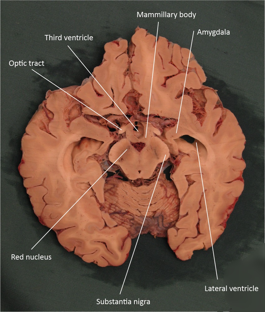
Brain anatomy: axial midbrain

Brain anatomy: axial midbrain


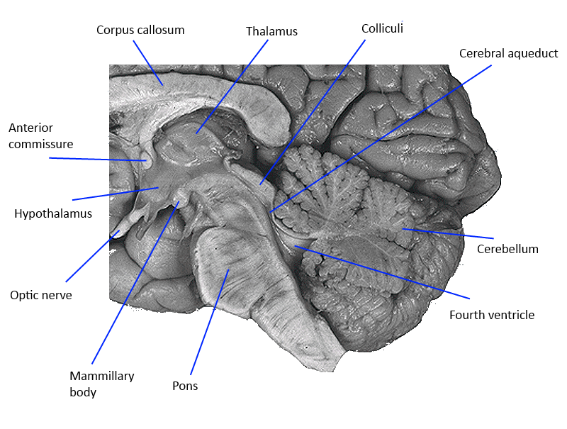
Brain anatomy: diencephalon

Brain anatomy: diencephalon


Brain and its blood supply

Brain and its blood supply


The body supplies blood to the brain via the internal carotid and vertebral arteries. These vessels come together to form a ring called the circle of Willis. The function of the circle of Willis is to provide a shunt system should any of the vessels become damaged.

Arising from the circle are the three main vessels that supply the brain with blood; the anterior cerebral artery, middle cerebral artery, and the posterior cerebral artery. 

It is important to be able to recognise the consequences of the occlusion of the three main vessels supplying the brain.

Anterior cerebral artery (ACA) occlusion

Occlusion of the anterior cerebral artery may result in the following defects

  • Hemiparesis of the contralateral foot and leg (more severely than the arm)
  • Sensory loss of the contralateral foot and leg
  • Motor dysphasia

If stroke occurs prior to the anterior communicating artery it is usually well tolerated secondary to collateral circulation

Middle cerebral artery (MCA) occlusion

Occlusion of the middle cerebral artery may result in the following defects

  • Hemiparesis of the contralateral face and limbs
  • Sensory loss of contralateral face and limbs
  • Dysphasia (when dominant hemisphere affected)
  • Contralateral neglect
  • Homonymous hemianopia or quadrantanopia
  • Dorsolateral prefrontal dysfunction

Posterior cerebral artery (PCA) occlusion

Occlusion of the posterior cerebral artery may result in the following defects

  • Alexia without agraphia (left PCA)
  • Contralateral loss of pain and temperature sensation
  • Contralateral hemianopia
  • Prosopagnosia
  • Ipsilateral cranial nerve defects (V, VIII, IX, X, & XI)
  • Horner's syndrome

Brain lesions

Brain lesions


The following neurological disorders/features may allow localisation of a brain lesion:

Gross anatomy

Parietal lobe lesions
  • sensory inattention
  • apraxias
  • astereognosis (tactile agnosia)
  • inferior homonymous quadrantanopia
  • Gerstmann's syndrome (lesion of dominant parietal): alexia, acalculia, finger agnosia and right-left disorientation

Occipital lobe lesions
  • homonymous hemianopia (with macula sparing)
  • cortical blindness
  • visual agnosia

Temporal lobe lesion
  • Wernicke's aphasia: this area 'forms' the speech before 'sending it' to Brocas area. Lesions result in word substituion, neologisms but speech remains fluent
  • superior homonymous quadrantanopia
  • auditory agnosia
  • prosopagnosia (difficulty recognising faces)

Frontal lobes lesions
  • expressive (Broca's) aphasia: located on the posterior aspect of the frontal lobe, in the inferior frontal gyrus. Speech is non-fluent, laboured, and halting
  • disinhibition
  • perseveration
  • anosmia
  • inability to generate a list

Cerebellum lesions
  • midline lesions: gait and truncal ataxia
  • hemisphere lesions: intention tremor, past pointing, dysdiadokinesis, nystagmus

More specific areas

AreaAssociated conditions
Medial thalamus and mammillary bodies of the hypothalamusWernicke and Korsakoff syndrome
Subthalamic nucleus of the basal gangliaHemiballism
Striatum (caudate nucleus) of the basal gangliaHuntington chorea
Substantia nigra of the basal gangliaParkinson's disease
AmygdalaKluver-Bucy syndrome (hypersexuality, hyperorality, hyperphagia, visual agnosia

Brain tumours

Brain tumours


The majority of adult tumours are supratentorial, where as the majority of childhood tumours are infratentorial.

Type of tumourFeatures
Gliolastoma multiforme• The most common primary brain tumour in adults.
• Histology: Pleomorphic tumour cells border necrotic areas
Meningioma• The second most common primary brain tumour in adults
• Histology: Spindle cells in concentric whorls and calcified psammoma bodies
Schwannoma• Often seen in the cerebellopontine angle: acoustic neuroma
• Bilateral schwannoms are seen in neurofibromatosis
• Histology: Antoni A or B patterns are seen. Verocay bodies (acellular areas surrounded by nuclear palisades)
Pilocytic astrocytoma• The most common primary brain tumour in children
• Histology: Rosenthal fibres (corkscrew eosinophilic bundle)
Medulloblastoma• More common in children
• Found exclusively in the posterior fossa
• Metatases through the CSF
• Histology: Small, blue cells. Rosette pattern of cells with many mitotic figures
Ependymoma• Commonly seen in the 4th ventricle
• May cause hydrocephalus
• Histology: perivascular pseudorosettes
Oligodendroma• Benign, slow-growing tumour common in the frontal lobes
• Histology: Calcifications with 'fried-egg' appearance
Haemangioblastoma• Vascular tumour of the cerebellum
• Associated with von Hippel-Lindau syndrome
• Histology: foam cells and high vascularity
Pituitary adenoma• Most common type is a prolactinoma
• May present with bitemporal hemianopia
Craniopharyngioma• Most common paediatric supratentorial tumour
• Histology: Derived from remnants of Rathke pouch
Metastases• Most common type of brain tumour

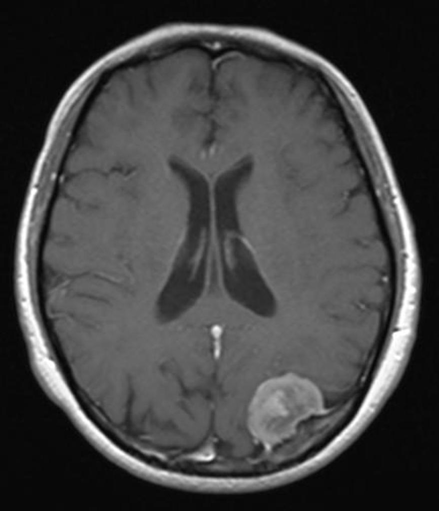
© Image used on license from Radiopaedia
Meningioma - MRI showing the typical well-circumscribed appearance. A dural tail can be where the tumour 'connects' to the dura. It is seen in around 65% of meningiomas.

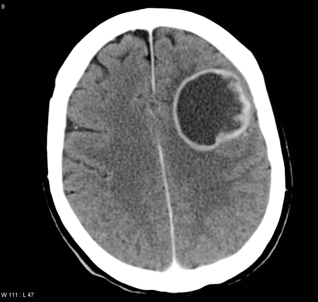
© Image used on license from Radiopaedia
Glioblastoma multiforme - CT showing a peripherally enhancing lesion within the left frontal lobe. Note the contrast to the more homogenous meningioma above.